
Emergency? Call 999 or go to the nearest emergency department. This website is not an emergency channel.
Minor surgery · Xanthelasma removal
Xanthelasma removal, after a proper assessment.
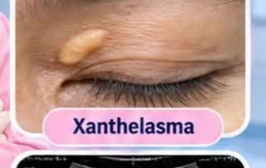
Xanthelasma are soft, yellowish plaques of cholesterol that form on or around the eyelids, most often at the inner corner of the eye. They are not dangerous in themselves. They are, however, worth taking seriously: a share of people who develop them also have raised blood cholesterol, and the eyelid is sometimes where that gets noticed first.
A consultation does not guarantee a procedure. A doctor assesses suitability, alternatives and whether you need a referral.
Not every eyelid lesion is xanthelasma. A doctor has to examine it. Do not use this page to diagnose yourself, and do not attempt removal at home.
01 · WHAT IT IS
Yellow deposits in the skin of the eyelid.
Xanthelasma (the singular form is xanthelasma; you will also see xanthoma used for the general family) is a collection of lipid-laden cells sitting in the upper layer of the skin. It appears as a soft, slightly raised, yellow-to-cream plaque. It is usually painless and does not itch.
The deposits are common around the inner corner of the upper eyelid, and can appear on all four lids. They are more frequent in middle age and beyond, and in people of Asian and Mediterranean descent.
Why the cholesterol link matters
Roughly half of people with xanthelasma have abnormal lipid levels. That is why a doctor will usually ask about your cardiovascular risk and may recommend a lipid profile blood test. Removing the plaque does not change the underlying lipid level, so the deposit can come back if the cause is not addressed.
This is the honest framing: the removal is cosmetic and functional, but the blood test is the part that can matter to your health.
02 · REMOVAL METHODS
Three approaches, and the choice depends on your lesion.
The right method depends on the size, thickness, depth and position of the deposit, and on how close it sits to the eyelid margin. A doctor decides after examining it. This is a summary of what is used, not a menu you can pick from.
01. Surgical excision
The deposit is cut out under local anaesthetic through a superficial dissection that avoids the deeper eyelid structures. It tends to give the most complete removal for thicker plaques and has the lowest recurrence rate of the three. It needs a wound that closes, so there is a scar, however fine.
02. Laser ablation
A laser vaporises the deposit with precision. It suits smaller or more superficial plaques and is good around delicate structures. It avoids stitches. It can leave a temporary colour change in the treated skin, and thicker lesions may need more than one session.
03. Radiofrequency
Controlled heat removes the plaque without stitches. Quick, with redness and mild swelling that settles over days. It suits plaques that are not too deep.
Sometimes a method is combined, and sometimes removal of a thicker lesion is planned together with an eyelid procedure. That is a clinical judgement, not a preference.
03 · WHAT TO EXPECT
Assessment first, then the procedure, then a review.
01. Assessment
The doctor examines the plaques, asks about your health and any family history of high cholesterol, and may arrange a lipid profile blood test.
02. Method and consent
You are told which method is proposed, why, what the scar and recovery are likely to be, and what the recurrence risk is. You decide.
03. The procedure
Performed under local anaesthetic, so the area is numb. Most removals are short and done as an outpatient visit.
04. Aftercare and review
You are given wound care instructions and a review point. Recurrence is possible, so follow-up matters.
04 · QUESTIONS WORTH ASKING
Bring these to the consultation.
Is this definitely xanthelasma, or could it be something else?
Do I need a lipid profile, and who follows up the result?
Which removal method are you recommending for my lesion, and why that one?
What will the scar look like, and how long until it settles?
How likely is it to come back, and what would you do if it does?
BRANCH AVAILABILITY
Confirm the branch before you travel.
Minor surgery is not necessarily available at every branch. Availability by branch has not been confirmed for this website.
State your area and the treatment you are asking about. Do not send sensitive medical details that have not been requested.
General medical information
This content is not a diagnosis, a prescription, or a substitute for a consultation. If your condition is serious or an emergency, call 999 or go to the nearest emergency department.
Content status: this page requires clinical review and a review date before publication.